Få mere at vide
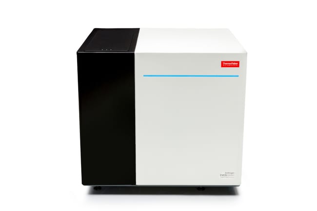
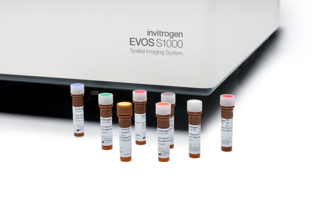
Invitrogen™ EVOS™ S1000 Rumligt billeddannelsessystem
Description
De spektrale egenskaber i EVOS S1000 billeddannelsessystemet muliggør samtidig indfangning og opløsning af op til otte fluorescensmål plus nuklear farvning (9-plex). Dette udføres i én enkelt erhvervelsesrunde, hvilket undgår behovet for gentagne mærknings- og billeddannelsescyklusser, der kan påvirke vævets integritet.
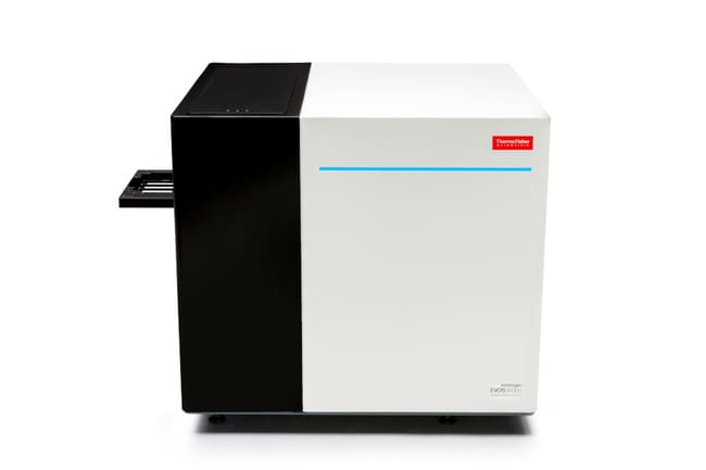
EVOS S1000-systemet har en fleksibel hardwarekonfiguration, der inkluderer en specialdesignet LED-baseret lysmotor og flere dikroiske og emissionsfiltre samt et højfølsomt sCMOS 4,2-megapixel kamera (6,5 m pixelstørrelse μ) til at opdage selv den mest indviklede vævsarkitektur. Systemet kan rumme op til fire slides og inkluderer tre forudindstillede mål (2,5X, 10X og 20X) med mulighed for at tilføje andre apokromatiske 5X- og 40X-objektiver.
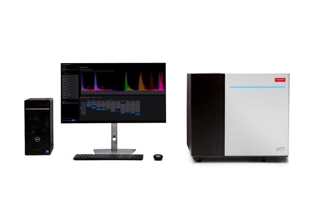
Instrumentet styres af EVOS S1000 rumsoftwaren, som tilbyder en sofistikeret, men enkel grænseflade til optagelse af hele vævsscanninger med integreret spektral unmixing. Softwaren tilbyder hurtige oversigtsscanninger i både transmitteret brightfield- og fluorescenstilstand samt 'periskop'-tilstand til levende vævsnavigation. Den tilbyder også en sofistikeret autofokusfunktion, der muliggør sammensyning og hele diasvævsscanninger, alt sammen problemfrit integreret for at undgå unødvendige processer efter erhvervelse eller offline.
EVOS S1000 Spatial Imaging System tilbyder disse vigtige fordele:
- Brugervenlig protokolbygger, der tillader nem udvælgelse af fluoroforer fra en dropdown-menu for automatisk at indstille den optiske konfiguration, instrumentet har brug for for at fange den ønskede plexitet
- Intuitiv autoeksponering og kraftfulde autofokus (laserbaserede) funktioner til hurtigt at lokalisere og identificere de ønskede mål
- Ligetil og konsekvent guidet arbejdsgang til spektralt opløsning (via lineær unmixing) op til 9-plex samples, ved brug af ufarvede og enkeltfarvede kontroller
- Færdiglavede og brugsklare protokoller til 9-plex og 7-plex billeddannelse, forudskabt fra referencevæv, som kan bruges til at starte billeddannelse af arkiverede vævsprøver med minimal indsats
- Bemærkelsesvis hurtig 9-plex optagelse af tilpasselige områdescanninger eller hele dias, hvor unmixing og stitching udføres automatisk som en del af billedoptagelsesprocessen
- Output af højkvalitets, OME-TIFF-billeder (med en opløsning på 325 nm/pixel ved 20X), som allerede er sammensyet, spektralt opløst og klar til brug med tredjepartssoftware til downstream-analyse, hvilket eliminerer behovet for mellemliggende justeringer
- Fuld kompatibilitet med fluorescerende farvestoffer fra flere leverandører, herunder Alexa Fluor, Alexa Fluor Plus, Aluorä Spatial Amplification og Opalä-farvestoffer, hvilket muliggør brug af enten primære antistoffluorescenskonjugater eller metoder til mærkning af rumlig forstærkning
- Fuld kompatibilitet med standard vævsdias og coverslips, og der kræves ingen proprietære forbrugsvarer
- Højfølsomt sCMOS-kamera (2040 x 2040 pixel opløsning, 4,2 megapixel) med 6,5 μm pixelstørrelse
Spektralbilleddannelse til rumlig biologi, gjort enkelt
EVOS S1000 Spatial Imaging System udnytter spektralbilleddannelsens kraft til samtidig at opløse 9 fluoroforer (8 proteinmål og DAPI) i en enkelt optagelsesrunde. Systemet er designet til at muliggøre farvevalg fra et udvalg af mere end 30 forskellige fluoroforer med emissionsspektre mellem 450 og 810 nm. Den anvender indbyggede spektralseparationsalgoritmer (baseret på lineær unmixing), som gør det muligt at identificere proteinmål klart og sikkert uden behov for iterativ mærkning og billeddannelse, hvilket hjælper med at bevare værdifulde vævsprøver.
Den fleksible spektralteknologi giver mulighed for at anvende og endda kombinere de vævsmærkningsmetoder, du ønsker, for succesfuldt at opdage mål med forskellige udtryksniveauer. Alle trin i anskaffelsesarbejdsgangen, der fører til et højkvalitets 9-plex billede, er designet til nem forståelse, selv for brugere, der måske er nye inden for rumlig billeddannelse og spektral unmixing.
Fremtidssikret og pålideligt hardware
EVOS S1000 Spatial Imaging System har en 4-positions præcisionsholderer på en motoriseret, softwarestyret, automatiseret scene. Instrumentet kan spektralt detektere mere end 60 forskellige kombinationer af excitation og emissionsbølgelængde, hvilket sikrer, at der ikke kræves yderligere filtre til fremtidige rumlige proteomiske billeddannelsesprojekter. Med en objektivkapacitet på 5 positioner kan forstørrelser tilpasses mellem 2,5X og 40X med apochromat-lignende objektiver.
Et højfølsomt sCMOS-kamera (2040 x 2040 pixel opløsning) bruges til at optage 16-bit monokrome OME-TIFF-billeder. Genererede billeder kan gemmes på den interne harddisk (2x 8-TB SSD'er til data), en ekstern USB enhed eller på kunde-IT-godkendte cloud-løsninger og lokale netværk. En ekstern Dell™ XE4 PC-computer med 12. generations Intel™ Core™ i9-12900-processor, 128 GB DDR4 RAM og NVIDIA™ Quadro RTX™ A4000 grafikkort bruges til at drive EVOS S1000 Spatial Imaging System.
Kraftfuld software designet til sofistikeret enkelhed
EVOS S1000 Spatial Software giver brugerne mulighed for at få adgang til multiplex-billeddannelse på væv på den simplest mulige måde. Erhvervelsesgrænsefladen er designet med stor vægt på brugervenlighed, selv for uerfarne brugere og dem, der er nye inden for spektral afblanding. Den starter med en ekstremt hurtig oversigtsscanning, der kan udføres enten i transmitteret eller fluorescerende tilstand. For eksempel kan fire dias scannes i fluorescerende tilstand, inklusive etiketterne, på ~180 sekunder.
Live ('periskop') tilstand er tilgængelig for hurtigt at navigere i vævet. At finde prøver er hurtigt og præcist ved hjælp af en kraftfuld kombination af specialdesignet IR-laserfokus og softwareautofokus. En guidet arbejdsgang gør det muligt at udføre processen med oprettelse af afblandingsmatrixen effektivt og sikkert, herunder udarbejdelse af en rapport om afblanding af kvalitetsmålinger med kvalitative og kvantitative parametre til at evaluere kvaliteten af den genererede spektrale afblandingsmatrix.
EVOS S1000 Spatial Software kan bruges til at køre variable forstørrelsesscanninger af FOV og ROI med flere muligheder for automatiserede rutiner. Hele slide-syning og -blanding udføres automatisk som en del af billedoptagelsen.
Parathed til downstream-analyse
Billeder optaget med EVOS S1000 Spatial Imaging System kan yderligere analyseres ved hjælp af enhver tredjepartssoftware, der understøtter pyramideformet OME-TIFF-filoutput til håndtering af store multidimensionelle billeddata. Eksempler er Halo-softwaren fra Indica Labs og den frit tilgængelige open source QuPath-analysesoftware.
EVOS S1000 Spatial Imaging System er bygget fra bunden for at gøre rumlig biologi, og især proteomisk vævsbilleddannelse, tilgængelig for ethvert laboratorium.
Specifications
Specifications
| Type | Konfigureret billedsystem |
| Produktlinje | EVOS |
| Kamera | 4,2 MP højt følsomt sCMOS-kamera |
| Til brug med (applikation) | Fluorescens, brightfield, fasekontrast og farveafbildning af vævspræparater |
| Til brug med (udstyr) | EVOS |
| High-throughput kompatibilitet | Nej |
| Lyskilde | Specialfremstillet LED-motor med følgende excitationsspektra: 375 nm, 405 nm, 440 nm, 500 nm, 530 nm, 570 nm, 630 nm og 730 nm |
| Mål | 5-positions automatiseret (inkluderer 2,5x, 10x og 20x) |
| Scene | Motoriseret |
| Dataoutput | Optagede billeder er standardiserede OME-TIFF filformatbilleder: rå fliser, ublandede eller syet og ublandede (hvis ublandede fliser er påført) |
| Show More |
Må kun benyttes til forskning.
By clicking Submit, you acknowledge that you may be contacted by Fisher Scientific in regards to the feedback you have provided in this form. We will not share your information for any other purposes. All contact information provided shall also be maintained in accordance with our Privacy Policy.